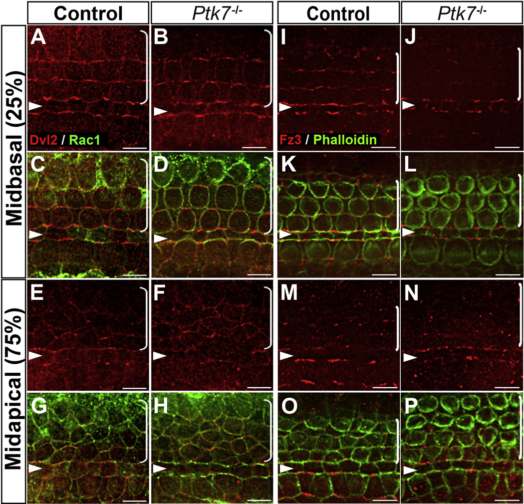
Figure 1. Ptk7 regulates Fz3 localization but is not required for asymmetric localization of Dvl2 in the OC.
(A–D) In the mid-basal region of the OC at E17.5, Dvl2 (red) was enriched on the lateral membranes of hair cells in both control (A, C) and Ptk7−/− cochleae (B, D). (E–H) In the mid-apical region of the OC at E17.5, similar membrane localization of Dvl2 was observed in control (E, G) and Ptk7−/− cochlea (F, H). Cell boundaries were labeled with Rac1 immunostaining (green). (I–L) In the mid-basal region of OC at E17.5, Fz3 (red) was enriched on the medial membranes of hair cells and supporting cells in control (I, K). This localization was disrupted in Ptk7−/− cochleae (J, L). (M–P) In the mid-apical region of the OC at E17.5, asymmetric localization of Fz3 was not apparent in either control (M, O) or Ptk7−/− cochleae (N, P). Green, phalloidin staining. Percentages indicate the distance of the positions analyzed from the base relative to the length of the cochlea. Arrowheads indicate the row of pillar cells. Brackets indicate OHC rows. Lateral is up in all micrographs. Scale bar: 6 µm. (See also Figure S1).

