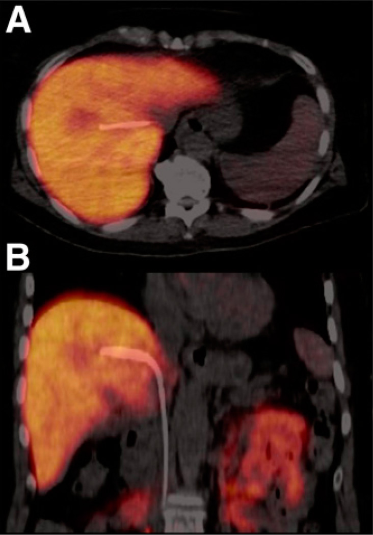
FIGURE 1.
Transaxial (A) and coronal (B) 18F-FDGal PET/CT images of mean tissue radioactivity concentration (kBq/mL liver tissue) from PET study without galactose infusion (subject 6). The position of the liver vein catheter inserted via the right femoral vein is seen. Note that the tracer accumulates specifically in liver tissue.

