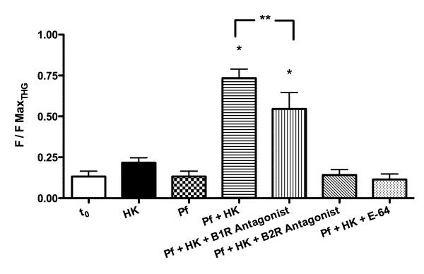
Figure 9.
Intracellular calcium mobilization in HUVECs cells after B1 and B2 receptors activation by kinin peptides. HUVECs were loaded with Fluo-3 AM and incubated with HK (20 μg) (HK); 10 μl of parasite culture supernatant without HK (Pf); 10 μl of parasite culture supernatant after incubation with 1.9 μg/μL of HK for 30 min. (Pf + HK); 10 μl of parasite culture supernatant after incubation with 1.9 μg/μL of HK for 30 min and pre-incubation (10 min.) with 4 μM Des-Arg9Leu8-BK (Pf + HK + B1 antagonist); or 10 μM HOE-140 (Pf + HK + B2 antagonist); 10 μl of parasite culture supernatant after incubation with 1.9 μg/μL of HK for 30 min (Pf + HK) pre-incubated with 5 μM E-64 (Pf + HK + E64). Data were normalized as the ratio between measured fluorescence (F) and maximal fluorescence (FmaxTHG) obtained with addition of 5 μM THG. *Denotes statistical significance with respect to all groups values p < 0.01 (n = 3; 30 cells); **Statistical significance of p < 0.05 between Pf + HK and Pf + HK + B2 antagonist (n = 3; 30 cells) in One-way ANOVA test and Newman-Keuls Multiple Comparison test.

