
Figure 1.
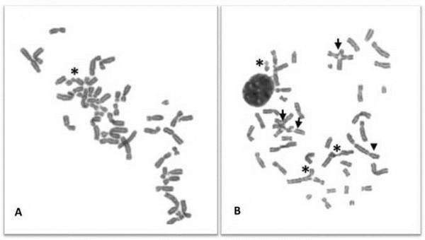
CI pattern in metaphases from DEB-induced lymphocyte cultures from a healthy donor and FA patient. Chromosomes were stained in a 4% Giemsa solution. The images were observed with an optical microscope (Olympus CX31) and captured with a digital camera (Nikon Sightds-smc), with the software DP20-5E microscope digital camera. (A) Selected metaphase from a HD lymphocyte culture exposed to 0.2 μg/ml of DEB for 48 h. One chromatid break can be seen (asterisk). (B) Selected metaphase from a FA patient lymphocyte culture exposed to 0.05 μg/ml of DEB for 48 h. High level of chromosome instability can be visualized, especially being important the tri and tetraradial figures (arrow) that are the hallmark for the diagnosis of FA. It can also be seen a dicentric chromosome (head arrow) and 3 chromatid breaks (asterisks).