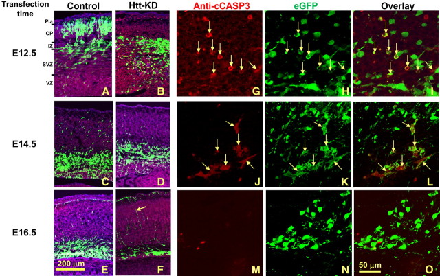
Figure 2.
The effect of Htt shRNA on cell migration and survival is time dependent in the developing cerebral cortex. A–O, Htt shRNA transfection was performed at E12.5 (A, B, G–I), E14.5 (C, D, J–L), and E16.5 (E, F, M–O) with 72 h survival. For E12.5 transfection, while many EGFP-labeled control cells migrate to the CP (A), Htt shRNA-transfected cells remain in the IZ (B). For E14.5 and E16.5 transfection, control transfected cells have left the VZ and largely reside in the IZ, with some cells present in the CP (C, E). Following Htt shRNA at these time points, most EGFP-labeled cells still remain in the VZ or the nearby SVZ (D, F). Pia, Pia mater. G–O, Higher-magnification confocal images show that cleaved CASP3-positive cells were also Htt shRNA positive and were easily detectible in the E12.5 condition (G–I, arrows). The apoptotic cells were greatly reduced at E14.5 (J–L, arrows), and only a few were observed at E16.5 transfection (M–O). Red, Anti-cleaved CASP3 immunostaining (anti-cCASP3); green, EGFP. Scale bars: (in E) A–F, 200 μm; (in O) G–O, 50 μm.

