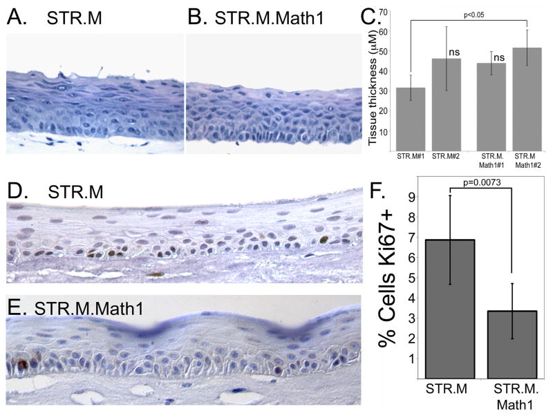
Figure 5. Math1 expression reduces STR cell proliferation but does not alter epithelial thickness when cells cultured under organotypic conditions.
Hematoxylin and eosin stain of epithelial tissues sections from A. STR.L control cells and B. STR.M.Math1 cells after cultured under organotypic conditions. C. Quantification of epithelial thickness, measured in three regions from three different tissue sections per culture. n=9. ns not significantly different by ANOVA and Tukey Rank Mean testing. D. and E. Ki67 staining in tissue sections from D. STR.M and E. STR.M.Math1 cells. F. Quantitation of Ki67 positive staining. The number of Ki67+ cells was expressed as a percentage of the total number of basal keratinocytes in a counted field. n=6

