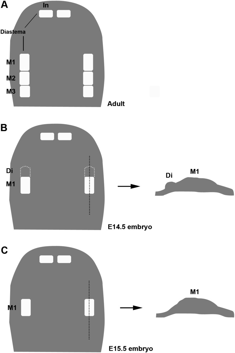
Figure 1.
Schematic diagrams showing tooth buds in rodent maxilla. Schematic representations of maxilla from wild-type mouse at adult (A), E14.5 (B), and E15.5 (C). Right diagrams of B and C showing sagittal view of tooth germs. In, incisor; M1, first molar; M2, second molar; M3, third molar; Di, rudimentary tooth bud.

