Figures 8–13.
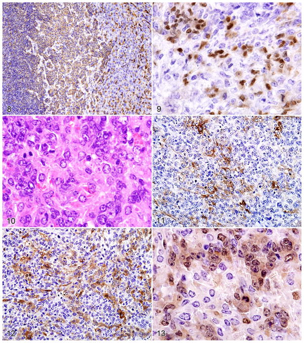
Figure 8. Histiocytic sarcoma in the liver stained with antibody to F4/80 showing high-level expression by normal Kupffer cells (right side), lower expression by the round–cell component of the histiocytic sarcoma (middle portion), and the lowest levels of expression by the spindle–cell component (left side). Figure 9. PAX5 staining of a spindle–type histiocytic sarcoma occupying the red pulp and surrounding a follicle with normal B cells. Note nuclear PAX5 staining of some spindle cells as well as normal B cells (upper left).
Histopathologic and immunohistochemical features of histiocyte-associated lymphoma. Serial sections of a histiocyte–associated lymphoma stained with HE showing the following: Figure 10. B-cell lymphoma and eosinophilic histiocytes. Figure 11. Antibodies to F4/80. Figure 12. Mac–2. Figure 13. PAX5. Note the more extensive staining of the histiocyte population by Mac–2 than F4/80 as they weave among the clusters of PAX5+ cells.

