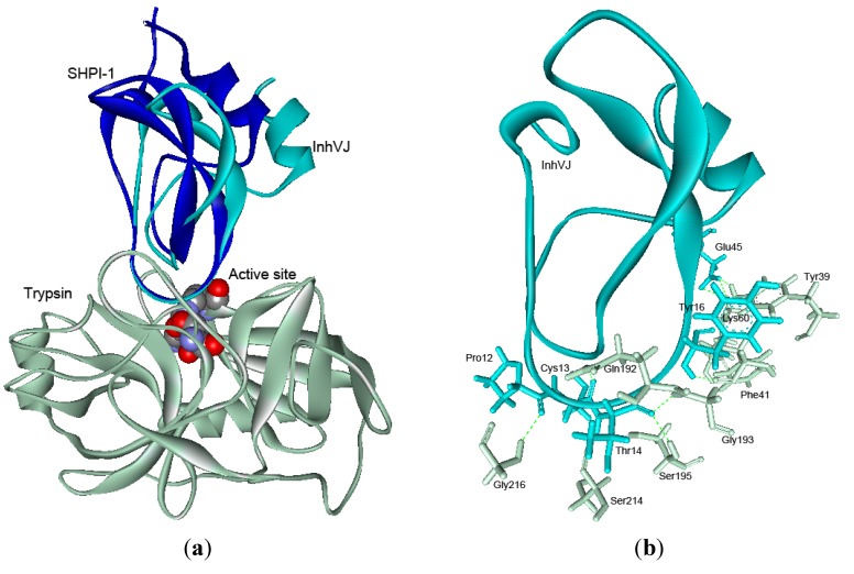
Figure 7.
(a) Superimposition of the theoretical model of complex InhVJ-trypsin and crystal structure of complex SHPI-1-trypsin (PDB ID 3M7Q). Structures are shown as the ribbon diagrams. The key residues of the enzyme active site are shown as bowls. (b) Intermolecular hydrogen bonds in InhVJ-trypsin complex and interacting amino acid residues are shown as sticks for InhVJ in cyan and for trypsin in light gray colors.

