
Fig. 2.
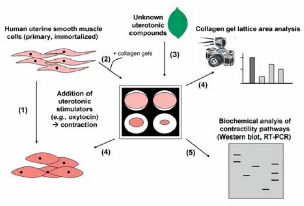
Overview of collagen gel uterine contractility assay. Human myometrial smooth muscle cell lines (e.g., telomerase-immortalised human myometrial cells or cell lines derived from a primary culture of human myometrial cells) contract in the presence of uterotonic stimulators, such as oxytocin (1). Collagen gels will be seeded in culture dishes with an appropriate amount of myometrial cells per well (based on the technique described by Fitzgibbon et al. [83]) (2). Cells in collagen gels will be allowed to equilibrate overnight in serum free medium. Various plant compounds of interest will be added to the serum free media and gels released from the culture dishes (3). The effect of collagen contractility will be monitored and quantified over time. The effect of concentration gradients of the various compounds on contractility will also be evaluated by this method (4). To determine which biochemical pathways are involved in the modulation of myometrial contractility various compounds that act as inhibitors of myometrial contractility pathways may be added to the collagen gel mix, prior to treatment of unknown plant compounds and gel release. Further investigations at the molecular biology level by isolating RNA and protein from the cells in collagen may be performed (5).