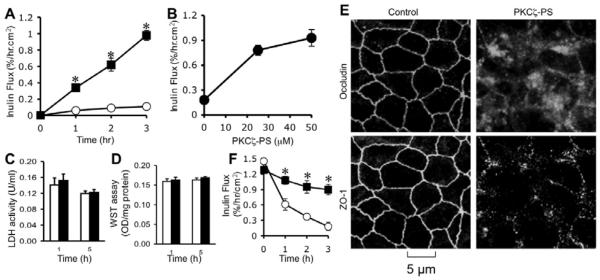
Fig. 3. Inhibition of PKCζ activity disrupts barrier function and delays tight junction assembly in MDCK cell monolayers.
A, B: MDCK cell monolayers were incubated with (■) or without (○) 50 μM PKCζ-PS for varying times (A) or with varying concentrations of PKCζ-PS for 2 hours (B). Barrier function was evaluated by measuring unidirectional flux of FITC-inulin. Values are mean ± s.e.m. (n = 6). Asterisks indicate the values that are significantly (P<0.05) different from corresponding control values. C, D: The cell viability assessed by measuring LDH activity in the incubation medium (C) or metabolic activity in the cell by WST assay (D) at 3 hour after 50 μM PKCζ-PS (black bars) or control peptide (white bars) administration. Values are mean ± sem (n = 6). E: MDCK cell monolayers incubated with or without 50 μM PKCζ-PS for 2 hr were fixed and stained for occludin and ZO-1 by immunofluorescence method. Images collected by confocal microscopy. F: Cell monolayers were incubated in low calcium medium for 16 hours to deplete extracellular calcium. Regular medium containing calcium with (■) or without (○) PKCζ-PS (10 μM) was then replaced. Inulin permeability (B) was measured at varying times after calcium replacement. Values are mean ± sem (n = 6). Asterisks indicate the values that are significantly (P<0.05) different from corresponding values for control cells.

