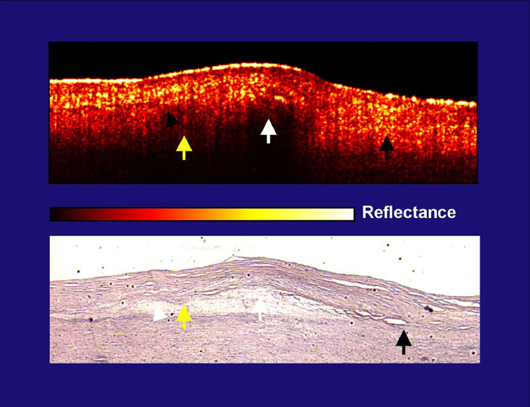
Figure 1. Lack of correlation in diffuse boundaries and lipid content.
This figure is from the initial Circulation publication from 19963. The yellow arrows demonstrate the cap-lipid interface that is diffuse and is covered by a highly scattering cap (yellow reflections in cap). The white arrow, which is also over lipid, identifies a cap with lower scattering and the cap-lipid interface is sharply defined. Both are over the same lipid collection. Finally, the black arrow shows the intimal-elastic layer interface (no lipid present) that is diffuse, with an intima that is highly scattering. In figure 1B, E, and F of the S-J Kang et. al. paper, there are diffuse border plaques, but the caps over them are highly scattering, which is potentially the etiology. The concern is therefore whether the lipid interface is diffuse in some OCT images because of the core composition, as others have previously suggested, or high scattering from the cap, as we have previously suggested. It is possible that highly scattering caps correlate with lipid plaques, but it is also possible that this scattering is the etiology for the variations in sensitivity and specificity. As clinical studies are now being performed in large numbers due to the availability of the technology, like the paper in this issue, OCT’s lipid characterizing ability needs to be more firmly established.

