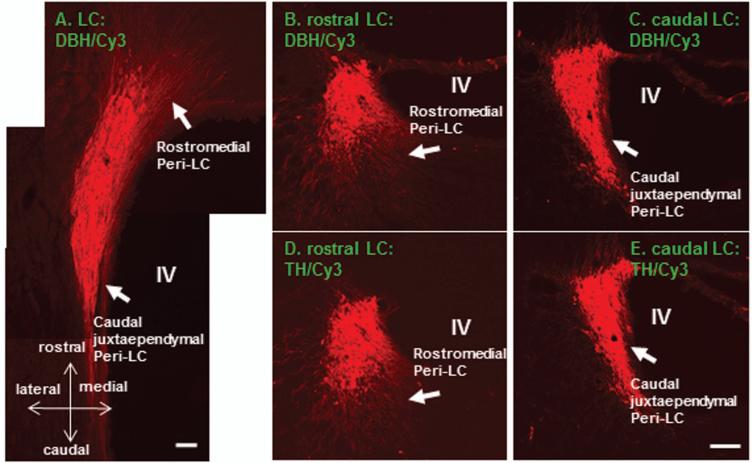
Figure 1.
Color photomicrographs illustrate DBH or TH immunoreactive profiles in the LC (A-E; Cy3: red). It is quite obvious that DBH and TH immunoreactivity in the adjacent sections revealing highly similar labeling patterns (B-E). Extranuclear LC processes are mainly noted in the rostromedial and caudal juxtaependymal Peri-LC. These two subregions are marked in B-E (arrows) and also are clearly visualized in the horizontal section (A). DBH: dopamine-beta-hydroxylase; TH: tyrosine hydroxylase; LC: locus coeruleus; IV: the IVth ventricle. Scale bar: 80 μm.

