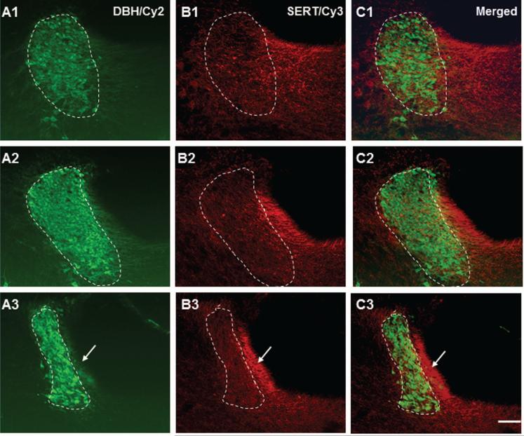
Figure 5.
Color photomicrographs demonstrate the spatial relationship between serotonin transporter (SERT) immunoreactive fibers and DBH positive LC nucleus (circled by dashed lines). The left column shows the LC from rostral to caudal (A1-A3) immunostained with DBH antiserum (Cy2: green). The middle column reveals SERT positive fibers from the same section (A2-C2; Cy3: red). The right column shows the merged photos. Note that SERT positive fibers are rather prevalent in the caudal juxtaependymal Peri-LC (marked by arrows in lower row). Scale bar: 60 μm.

