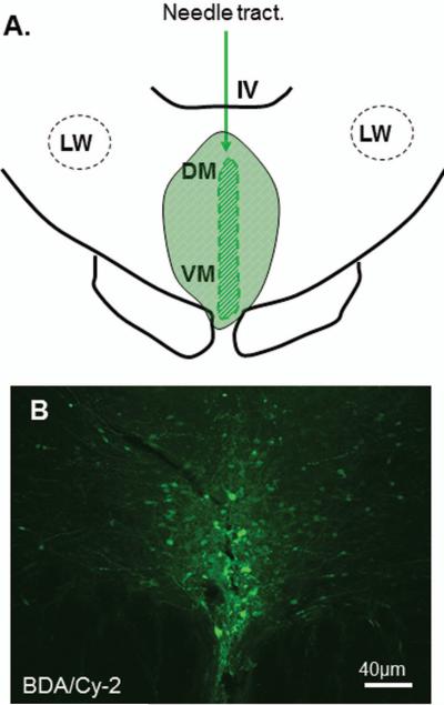
Figure 6.
Line drawing for the reconstruction of anterograde tracer injection site in the DR is shown in A. The fluorescent stained BDA (Cy2: green) reaction reveals the injection site in the DR (B). Note that a majority of BDA labeled cells are restricted in the DR. IV: 4th ventricle; LW: lateral wing; DM: dorsomedial; VM: ventromedial.

