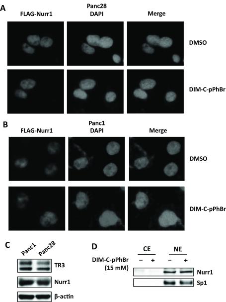
Figure 2.
Expression and subcellular localization of NR4A2 in Panc28 and Panc1 cells. Panc28 (A) and Panc1 (B) cells were transfected with adenovirus expressing FLAG-Nurr1 for 4 hr; media was changed and after 18 hr, cells were treated with 15 μM of DIM-C-pPhBr for 12 hr and immunostained with anti-FLAG antibody. Fluorescent images were obtained as described in the Materials and Methods. (C) Whole cell lysates from Panc1 and Panc28 cells were analyzed by western blotting and β-actin was used as a loading control. (D) Cells were treated with DMSO or 15 μM DIM-C-pPhBr for 24 hr, and cytosolic (CE) or nuclear (NE) extracts were analyzed by western blots (Sp1 was a nuclear protein control).

