
Fig. 1. Pdx1 autoantibodies in prediabetic NOD mice.
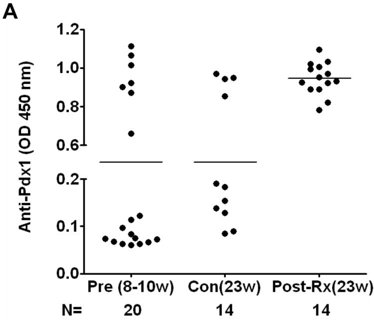
A. Detection of Pdx1 antibodies in NOD mice. Sera were collected from female NOD mice at various ages ranging from 8 to 23 weeks or NOD mice treated with daily injection of rPdx1 for 12 weeks. PAA were assayed using ELISA at a serum dilution of 1:30. Positive value is defined 3 SD above the mean of the BALB/c control sera. (Pre, pre-treatment; Con, PBS-treated controls; Post-Rx, treated with rPdx1).
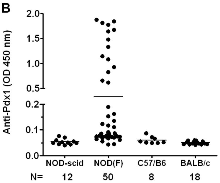
B. Comparison of PAA in various strains of mice. Sera from various mouse strains at different ages were collected and PAA were measured by ELISA and expressed as OD values at 450 nM using the 99th percentile of BALB/c control mice as the threshold definition of positivity.
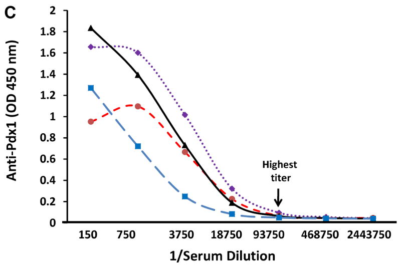
C. PAA titration curve. Selected PAA-positive serum samples at age of 10 weeks were serially diluted as indicated and OD values were determined by ELISA. Titer is defined as the last dilution giving positive OD reading above control.
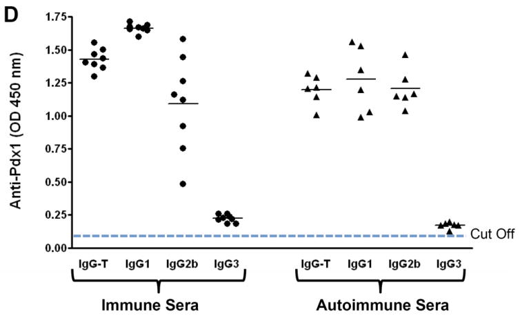
D. Isotypes of anti-Pdx1 antibodies. Total anti-Pdx1 antibody activity (IgG-T) and anti-Pdx1 antibodies of the IgG1, IgG2b, and IgG3 isotypes in sera from NOD mice immunized with Pdx1 (immune sera, solid circles) and selected PAA-positive autoimmune sera (triangle) from NOD mice were assayed by ELISA (1:30 dilution). An OD of 0.10 was used as the positive cut-off value (≥ 3 SD above the mean of the BALB/c control sera).