
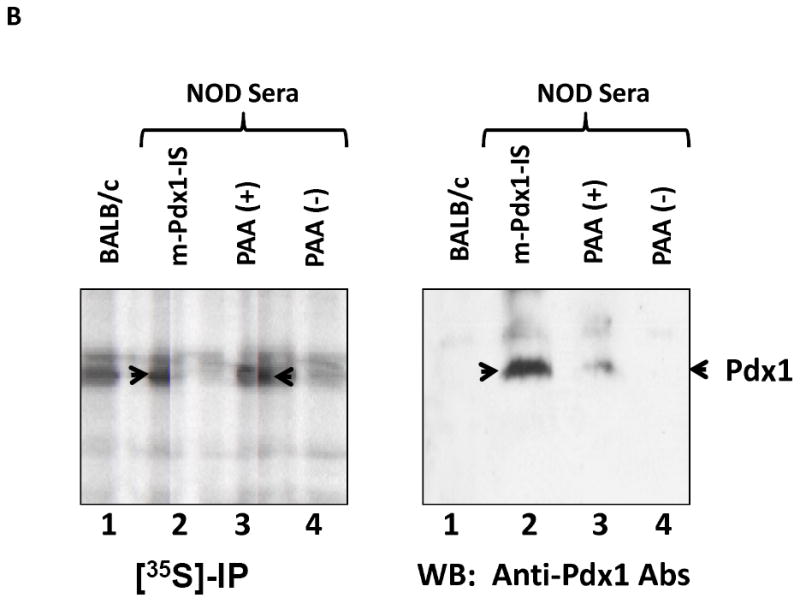
Fig. 2. Confirmation of PAA by Western blot (A) and immpreprecipitation (B).
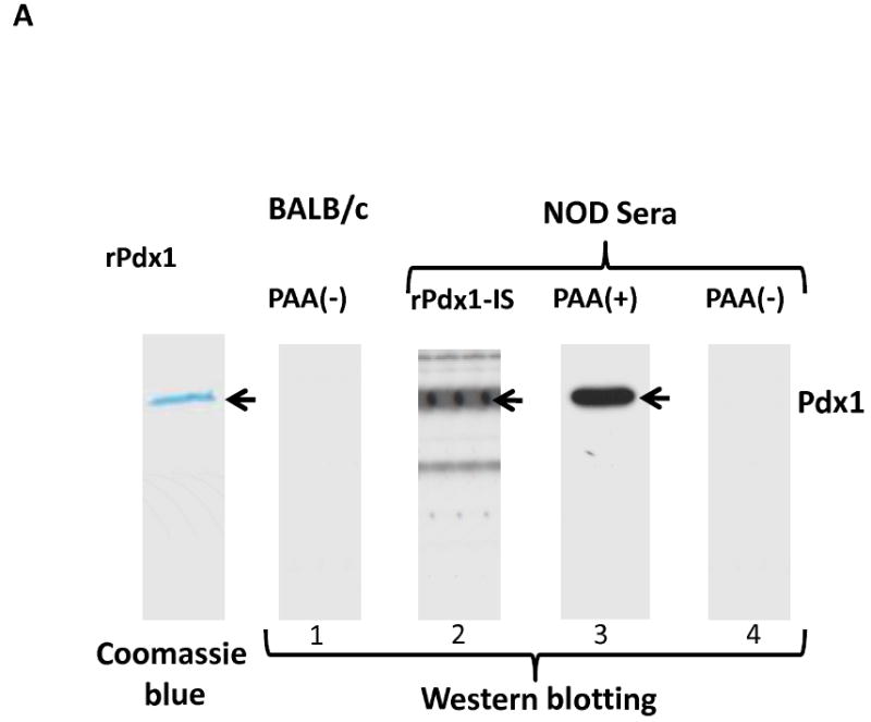
A. Western blotting. Purified recombinant rat Pdx1 protein (1 μg/lane) was separated on a 10% SDS-PAGE and stained with Coomassie blue or transferred to PVDF membrane. The membrane was probed with BALB/c or NOD mouse serum either positive (PAA+) or negative (PAA-) by ELISA, or with rabbit polyclonal anti-Pdx1 immune serum (rPdx1-IS) as positive control.
B. Immunoprecipitation and Western blotting. Rat insulinoma cells (INS-1) were labeled with [35S]-methionine overnight and cell lysate (0.5 mg) was incubated for 1 hr at 4°C with preformed immune complexes by incubating 10 μl mouse serum with 50 μl protein A/G overnight at 4°C. 35S-labelled proteins were separated by SDS-PAGE, fluorographed, and the dried gel was exposed to X-ray film at -80°C for 7 days. In parallel, unlabeled INS-1 cell lysate was subjected to immunoprecipitation with the same mouse sera. The immune complexes were separated by SDS-PAGE, transferred to PVDF membrane, and probed with rabbit anti-Pdx1 polyclonal antibodies (1:2000). Lanes 1-BALB/c, 2- Pdx1-treated NOD mouse immune serum (m-Pdx1-IS), 3-PAA(+), and 4-PAA(-) NOD serum samples. Arrow indicates position of Pdx1 protein.