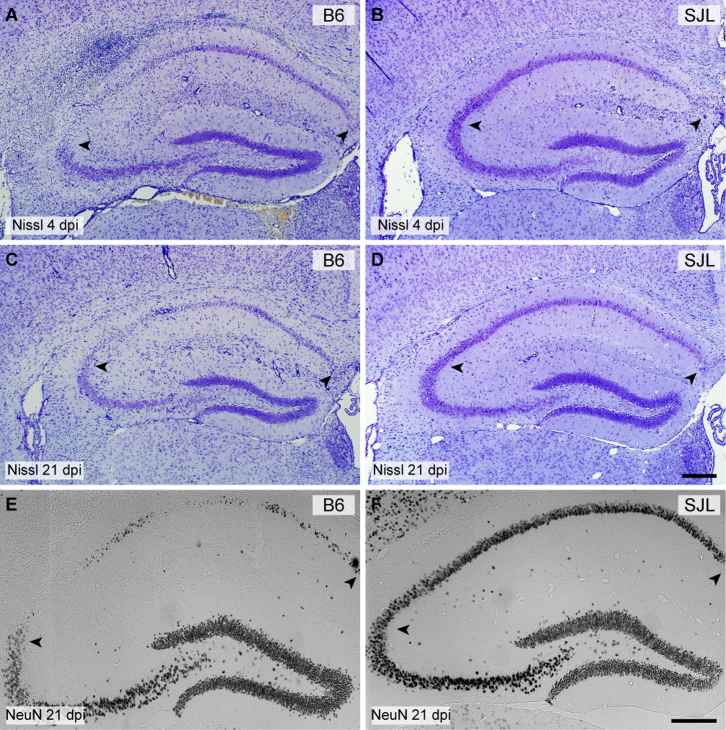
Figure 1. Injury to CA1 hippocampal neurons associated with acute TMEV infection is absent in SJL mice.
Hippocampal sections from B6 (A, C, E) and SJL (B, D, F) mice were processed for Nissl histochemistry (A–D) or immunostained for the neuron-specific marker NeuN (E–F) at 4 days postinfection (dpi) (A, B) or 21 dpi (C–F). By 4 dpi about 50% of CA1 region neurons are clearly lost in the B6 mouse (A) but minimal neuronal dropout is evident in the SJL mouse at this timepoint, despite the presence of a significant inflammatory response within the hippocampal fissure (B). By 21 dpi the majority of CA1 neurons are lost in B6 mice (C, E) while virtually all CA1 neurons are preserved in SJL mice (D, F). Scale bar in D is 250 μm and refers to A–C. Scale bar in F is 250 μm and refers to E. Arrowheads delineate the CA1 region. Sections are representative of at least 10 mice of each strain across multiple (>3) experiments.

