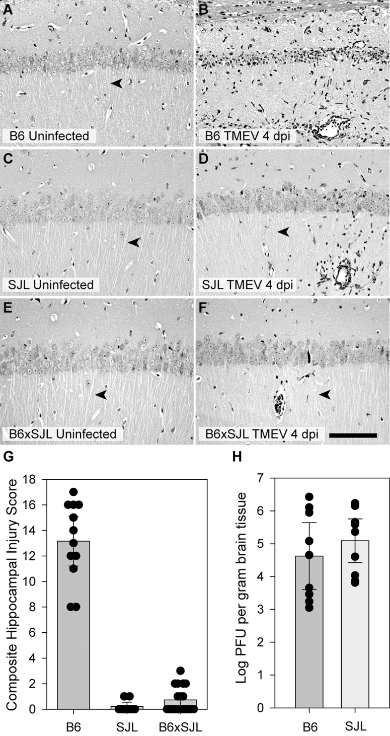
Figure 2. Hippocampal protection during acute TMEV infection is genetically dominant and viral replication does not explain the protection of CA1 neurons in SJL mice.
Hippocampal sections from uninfected (A, C, E) and 7 dpi (B, D, F) B6 (A, B), SJL (C,D), and B6xSJL F1 hybrids (E, F) were stained with hematoxylin and eosin. F1 hybrids, like the SJL parentals, do not exhibit substantial hippocampal injury. The extent of hippocampal injury was quantified (G) by summing the estimated percentage of CA1 neurons lost in each hemisphere represented on a scale of 0 to 10 (for example, the injury in Figure 1A is a 5). Zero indicates no injury to either hippocampus, while 20 represents complete loss of CA1 in both hemispheres. While there was no difference between SJL and F1 hybrid mice (B6xSJL), the injury observed in C57BL/6 mice was highly significant (P<0.001) compared to both groups. Viral load in the brain at 7 dpi was measured by standard plaque assay using brain homogenates incubated on L2 cells for 72 hours. (H) No difference was observed between B6 and SJL mice. Measurements for individual animals are shown in the scatter plots. The bar graphs represent the mean and 95% confidence intervals (95% CI) for the groups. Scale bar in F is 100 μm and refers to all panels. Sections are representative of at least 10 mice from each group in 3 separate experiments.

