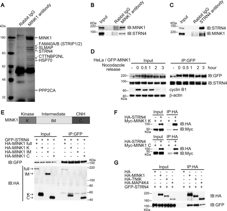
FIGURE 4.
MINK1 associates with STRN4. A, 293T cells were lysed, and the lysates were immunoprecipitated with anti-MINK1 antibody. The immunoprecipitates were separated by SDS-PAGE gel and silver stained. B, 293T cells were lysed, and the lysates were immunoprecipitated with either control or anti-MINK1 antibody; the immunoprecipitates were blotted with anti-MINK1 or anti-STRN4 antibodies. C, 293T cells were lysed, and the lysates were immunoprecipitated with either control or anti-STRN4 antibody; the immunoprecipitates were blotted with anti-MINK1 or anti-STRN4 antibodies. D, GFP-MINK1 HeLa cells were nocodazole arrested and released. The cells were lysed at the indicated time points, and the lysates were immunoprecipitated with anti-GFP antibody; the immunoprecipitates were subjected to immunoblot with the indicated antibodies. E, GFP-STRN4 was transiently expressed in 293T cells with HA-tagged deletion constructs of MINK1. The cells were lysed, and the lysates were immunoprecipitated with anti-GFP antibody; the immunoprecipitates were blotted with anti-HA and anti-GFP antibodies. F, HA-STRN4 and Myc-tagged deletion constructs of MINK1 were translated in vitro. The proteins were mixed and then immunoprecipitated with anti-HA antibody; the immunoprecipitates were immunoblotted with anti-HA and anti-Myc antibodies. G, GFP-STRN4 was transiently expressed in 293T cells with HA-MINK1, HA-TNIK, and HA-MAP4K4. The cells were lysed, and the lysates were immunoprecipitated with anti-HA antibody; the immunoprecipitates were immunoblotted with anti-HA and anti-GFP antibodies.

