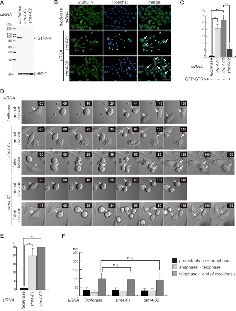
FIGURE 5.
STRN4 is required for abscission. A, HeLa cells were transfected with the indicated siRNA, and 72 h later, the cells were lysed and blotted with anti-STRN4 antibody. B, HeLa cells were cultured on glass coverslips and transfected with the indicated siRNAs. Seventy-two hours later, the cells were fixed and immunostained with anti-α-tubulin antibody and Hoechst. The arrows indicate multinucleated cells. C, HeLa cells were treated as in B, and the ratio of multinucleated cells was evaluated. To perform rescue experiment, HeLa cells were transfected with expression plasmid encoding GFP-STRN4 together with STRN4 siRNA (strn4–02) that targeted 3′-UTR of STRN4. Seventy-two hours later, ratio of multinucleated cells in GFP-positive cells were evaluated. Three independent experiments were performed, and 200 cells were evaluated for each experiment (the data are represented as the mean ± S.D.; **, p < 0.01). D, HeLa cells were transfected with control or STRN4 siRNAs; 48 h later, the cells were observed using time-lapse microscopy. Representative images of two types of cell divisions of STRN4 siRNA-transfected cells are shown. normal division indicates cells that completed cytokinesis. failed division indicates cells that were unable to complete cytokinesis. The arrows point dividing cells. E, graph shows the ratio of cells that failed cytokinesis. Forty cells for each group of siRNA-transfected cells were evaluated, and three independent experiments were performed (the data are represented as the mean ± S.D.; **, p < 0.01). F, time period of each stage of mitosis was evaluated. The graph shows the average time period of 30 cells for each group of siRNA-transfected cells (the data are represented as the mean ± S.D.; n.s., not significant).

