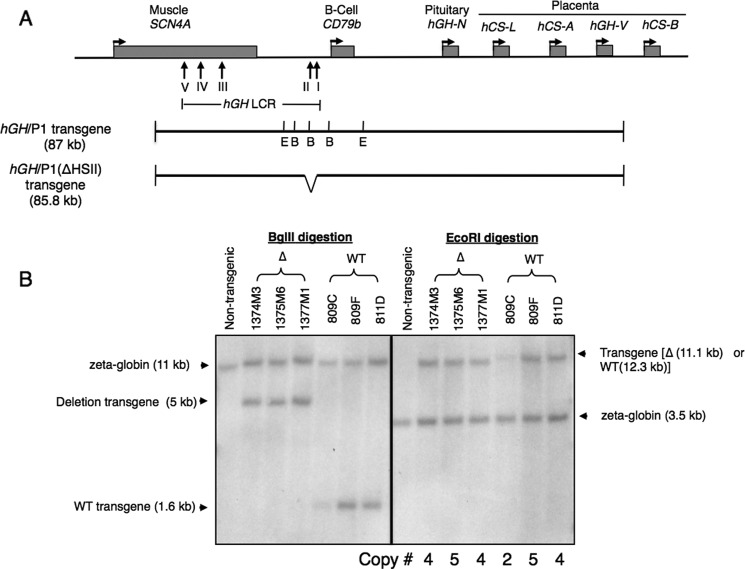
FIGURE 1.
Verification of hGH/P1(ΔHSII) transgene structure and determination of transgene copy number. A, map of the hGH locus and surrounding region on chromosome 17q22-23. The locations of the SCN4A and CD79b genes are shown. The vertical arrows indicate the positions of the DNase I hypersensitive sites of the hGH LCR. Maps of the intact hGH/P1 and hGH/P1(ΔHSII) constructs are shown below the map of the locus. The BglII (B) and EcoRI (E) sites used to confirm the target deletion and determine the copy numbers of the transgene are labeled (short vertical lines). The position of the 1.2-kb deletion is shown in the map of hGH/P1(ΔHSII). B, Southern blot analysis to confirm the targeted HSII deletion and determine the copy numbers of the transgene in each line. Genomic DNA from three hGH/P1 (WT) and three HSII deletion lines (Δ) was digested with BglII or EcoRI, run on an agarose gel, transferred, and hybridized with radiolabeled probes corresponding to the HSI region and to the endogenous mouse ζ-globin gene. Fragments resulting from the constructs and probes are indicated with arrows on the right-hand side of the image. A total of five digestion/copy number analyses were conducted. The 1.2-kb deletion resulted in loss of one BglII site 5′ to the HSII. Therefore, the 1.6-kb BglII fragment in the WT hGH/P1 is shifted to 5 kb in the hGH/P1(ΔHSII) transgenic pituitaries. The three previous described WT hGH/P1 lines were used as references to determine the copy numbers of the transgene (3), as indicated below the Southern blot.

