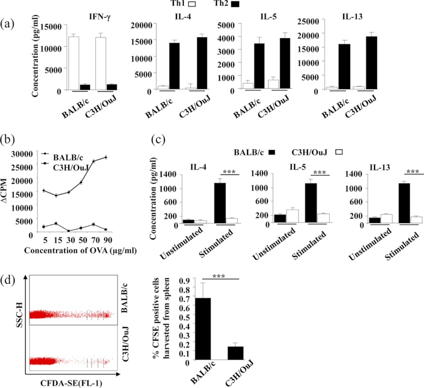
FIGURE 1.
BALB/c mice support survival of Th2 cells in vivo, whereas C3H/OuJ mice do not. a, BALB/c and C3H/OuJ mice generate comparable Th1 and Th2 cell responses in vitro. CD4+ T cells from BALB/c and C3H/OuJ strains were stimulated with plate-bound anti-CD3 (1 μg/ml) and anti-CD28 (2 μg/ml) antibodies under Th1- or Th2-priming conditions. Terminally differentiated cells were restimulated with plate-bound anti-CD3 and anti-CD28 antibodies, and supernatant was collected after 48 h for cytokine measurements. b, C3H/OuJ mice generate attenuated antigen-specific T cell responses to OVA immunization. Draining lymph node cells were harvested from mice immunized with OVA + alum and in vitro challenged with OVA at various concentrations. Proliferation was assessed following culture for 72 h in vitro and pulsing with [3H]thymidine for the last 8–10 h of incubation time. c, cytokine production was determined from the culture supernatants of the cells stimulated with 70 μg/ml OVA from the above culture plates after 48 h of incubation. d, OVA antigen-specific Th cells of both BALB/c and C3H/OuJ mice were expanded in vitro, CFDA-SE-labeled, and adoptively transferred in syngeneic BALB/c and C3H/OuJ mice (immunized with OVA + alum), respectively. After 5 days, spleens and lymph node cells were harvested and assessed for the presence and proliferation of the transferred cells using FACS. SSC-H, side scatter pulse height; CFSE, carboxyfluorescein succinimidyl ester. Representative data (mean ± S.E.) in triplicate wells from at least three independent experiments are shown. ***, p < 0.001 versus untreated.

