FIGURE 4.

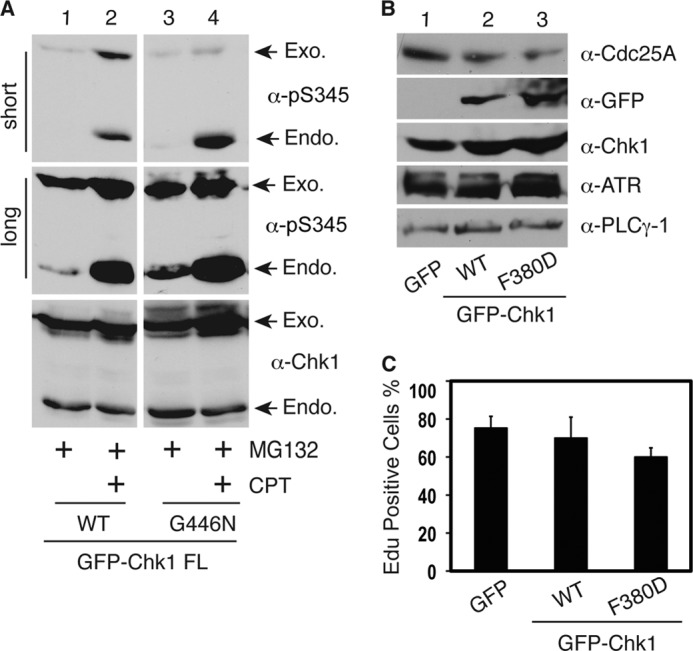
Cytoplasmic Chk1 mutants retain checkpoint function. A, HEK293T cells were transfected with GFP-tagged vectors expressing WT Chk1 or the G446N mutant for 48 h, treated or not with the proteasome inhibitor MG132 (2 μm) for 12 h, treated or not with 500 nm CPT for an additional 2 h, and immunoblotted with the indicated antibodies. Short and long exposure results for the anti-phospho-Ser-345 antibodies are shown. Exogenous (Exo.) and endogenous (Endo.) Chk1 proteins are indicated by arrows. B, HEK293T cells were transfected with vectors expressing GFP, GFP-WT Chk1, or GFP-F380D for 48 h and immunoblotted with the indicated antibodies. α-PLCγ-1, anti-phospholipase Cγ1 antibody. C, HeLa cells plated on glass coverslips were transfected with GFP, GFP-WT Chk1, or GFP-F380D for 24 h; synchronized at G2/M phase by 100 ng/ml nocodazole treatment for 20 h; washed; and released into fresh medium. After 16 h, cells were labeled with 10 μm 5-ethynyl-2′-deoxyuridine (EdU; a nucleotide analog) for 20 min and fixed, and 5-ethynyl-2′-deoxyuridine-positive cells were counted under a fluorescence microscope. Data represent the mean ± S.D. from three independent experiments.
