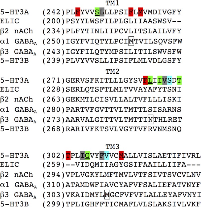
FIGURE 8.
Alignment of amino acid sequences of the TM1, TM2, and TM3 in the human 5-HT3A, ELIC, human β2 nAChR, human α1 GABAAR, human β3 GABAAR, and human 5-HT3B subunits. The position numbers of the first residue in the respective sequences are shown in parentheses to the left of the sequences. Green, residues in 5-HT3A where at least one of the introduced mutations had a significant effect on PU02 activity. Cyan, residues in 5-HT3A where mutations had a weak effect on PU02 activity. Red, residues in 5-HT3A where none of the introduced mutations had an effect on PU02 activity. Gray, residues in 5-HT3A where all introduced mutations resulted in “non-functional” 5-HT3A receptors. Underlined, residues in 5-HT3A where introduced mutations were combined into double mutants. Boxed, residues in α1 and β3 GABAAR subunits reported to be important for etomidate activity.

