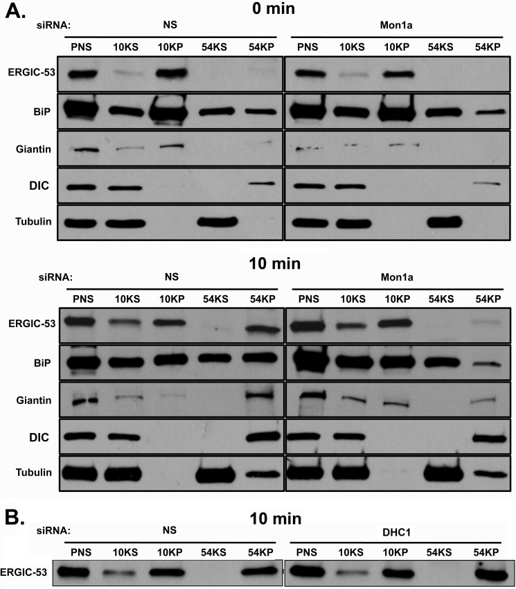
FIGURE 8.
Silencing of Mon1a delays formation of ERGIC-53-positive vesicles off the ER. A, NIH3T3 cells nonspecifically (NS) or Mon1a-silenced were treated with BFA as in Fig. 1. BFA was removed, cells were incubated for 10 min at 37 °C and homogenized, homogenates were centrifuged at 800 × g for 5 min (PNS), supernatant was centrifuged at 6,552 × g, 30 min (10KS and 10KP) to obtain an ER membrane fraction, and the remaining supernatant was centrifuged at 191,065 × g for 30 min (54KS and 54KP) to obtain ER-derived vesicles. Supernatants and membranes were solubilized in lysis buffer and resolved on SDS-PAGE, and ERGIC-53, BiP, Giantin, DIC, and tubulin localization was determined by Western blot analysis. A representative blot is shown (n = 3). B, cells as in A were nonspecifically or DHC1-silenced and were treated as in A, and ERGIC-53 movement into high-speed vesicles pellets was assessed. A representative blot of the 10 min recovery is shown (n = 3).

