Fig. 1.
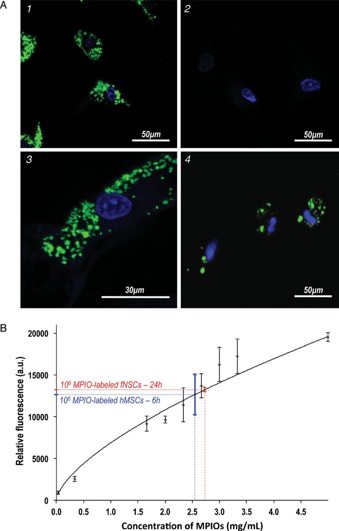
MPIO labeling of hMSCs and fNSCs. (A) Confocal fluorescence microscopy was performed on (1) MPIO-labeled hMSCs (60×), (2) unlabeled hMSCs, (3) a MPIO-labeled hMS cell (100×) and (4) MPIO-labeled fNSCs (60×). These results confirm the cytoplasmic localization of MPIO (green, Dragon green fluorescent tag) around the nuclei (blue, Hoescht staining) for both SC sources. (B) Level of fluorescence measured from a range of 9 concentrations of free MPIO in 200 μL of PBS (black, n = 4 per data point) and from 1 × 10⁶ MPIO-labeled fNSCs (red, n = 4) and 1 × 10⁶ MPIO-labeled hMSCs (blue, n = 4). Incubation times of 24 h for fNSCs and 6 h for hMSCs lead to a comparable level of fluorescence (P = .7).

