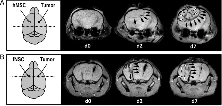
Fig. 2.
Tropism of MPIO-labeled hMSCS and fNSCs toward U87vIII tumors following contralateral injections. Axial T2*-w MRI slices of U87vIII tumor-bearing mice on the day of (d0), 2 days (d2), and 7 days (d7) after contralateral injection of (A) MPIO-labeled hMSCs and (B) MPIO-labeled fNSCs. The MPIO-labeled SC injection sites are shown as empty circles, the tumor injection sites as filled circles. The dotted lines show the localization of the displayed imaging slices. MPIO-labeled SCs localized at the edges of the tumor (black arrows) and inside the tumor masses (white arrows).

