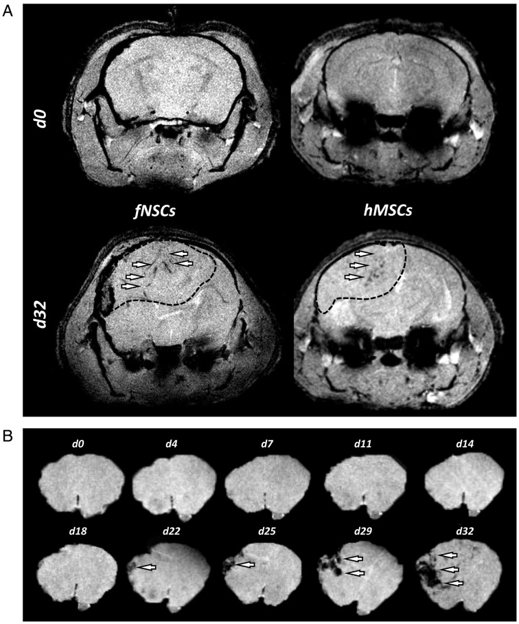
Fig. 5.
Biodistribution of MPIO-labeled hMSCS and fNSCs in GBM26 tumors following intratumoral injections. (A) Axial T2*-w MR images of GBM26 tumor-bearing mice on the day of (d0) and 32 days (d32) after intratumoral injection of MPIO-labeled hMSCS (left) and fNSCs (right). The dotted line shows the localization of the GBM26 tumor. (B) T2*-w axial images of a GBM26 tumor-bearing mice injected with MPIO-labeled hMSCs showing the brain region at days 0/4/7/11/14/18/22/25/29/32. MPIO-labeled SCs localized inside the tumor masses (white arrows).

