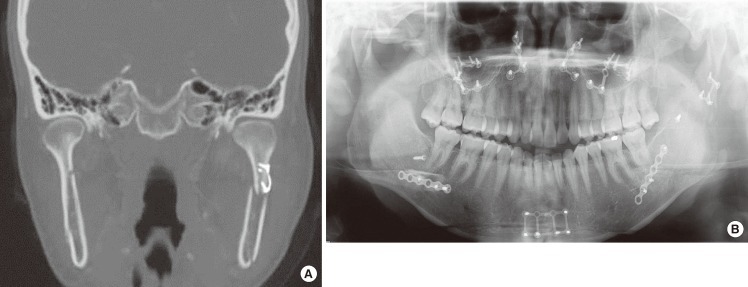
Fig. 9.
A 24-year-old woman with iatrogenic condyle fracture during orthognatic surgery
Open reduction and internal fixation was done. (A) Axial view of 3D head computed tomography (CT). (B) Panorama plain film. 3D head CT and panorama was checked at postoperative 7 weeks. The distal fracture segment was displaced medially and bone gap was checked 1.84 mm. In this case, the non-union or malunion will be occurred.

