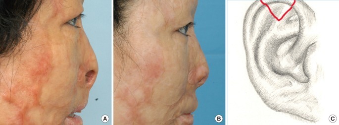
Fig. 2.
Case 2
(A) A 47-year-old female with both alar defects after a burn (Table 1, patient 11). Preoperative view shows alar defect. (B) 9×16 mm and 10×18 mm chondrocutaneous composite grafts from the helix were harvested and grafted. A frontal view 6 months after the operation shows improvement. (C) The donor site of the ear in this patient.

