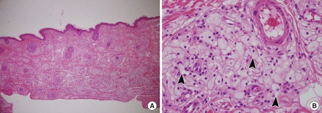
Fig. 4.
Histopathologic findings of the left upper eyelid lesion. (A) Pale areas containing foamy cells are dispersed throughout the dermis (H&E, ×40). (B) Xanthoma cells. These foamy histiocytes are polygonal or rounded with a distinct cell membrane. Their nuclei were small and eccentric and their cytoplasms are stuffed with lipid vacuoles (black arrowheads) (H&E, ×400).

