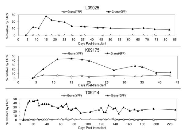
Figure 4. Percent GFP+ and YFP+ granulocytes in each animal by FACS analysis over the course of the study.
In each case, GFP+ granulocytes reach a peak within the first month post-transplant, and then decline and stabilize. YFP+ granulocytes, though present at very low levels, were detectable at each time point, even in L09025.

