FIGURE 1:

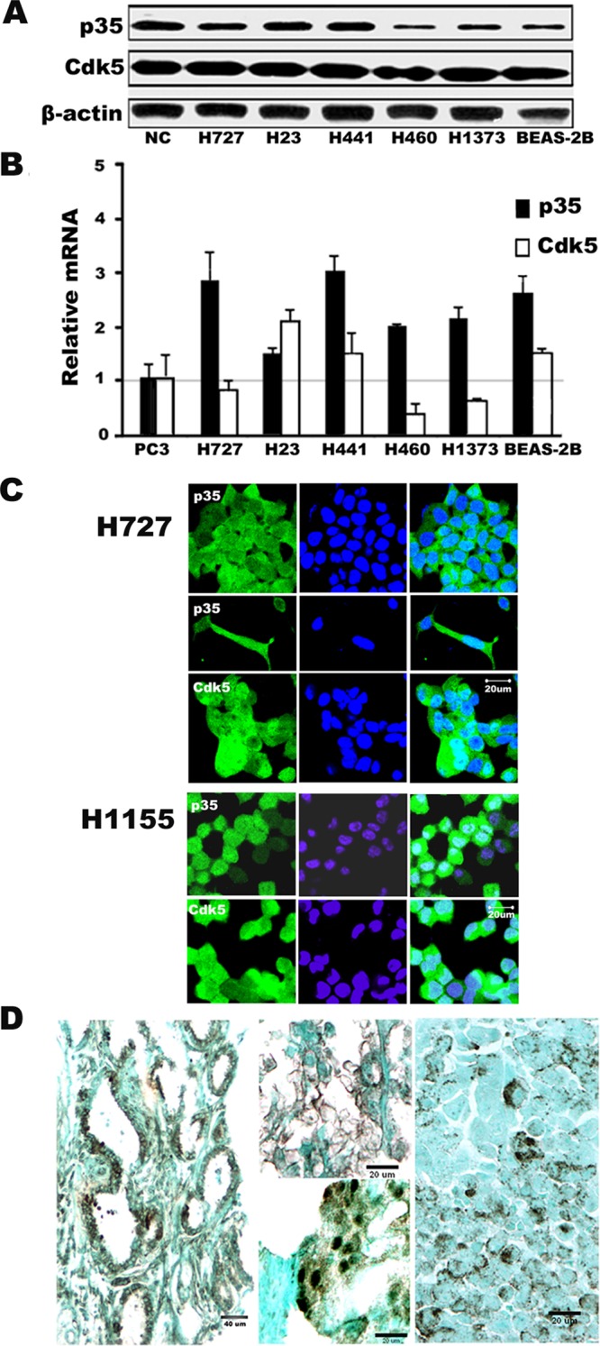
Expression of Cdk5 and the Cdk5 activator p35 in human lung cancer. (A) Western blotting of lung cancer (center lanes) and bronchial epithelial (BEAS-2B) cells shows expression of both p35 and Cdk5. NCs from mouse brain were used as a positive control (NC). (B) Relative mRNA expression by qRT- PCR shows high levels of p35 in all cell lines, while Cdk5 levels are variable. Expression is shown relative to the prostate cancer PC3 cells, which are designated as “1.” Mean ± SD from at least three independent experiments. (C) Photomicrographs of immunofluorescence (green) in two human lung cancer cell lines (H727 and H1155) reveal both cytoplasmic and nuclear distribution of p35 and Cdk5 protein. Nuclei were counterstained with DAPI (blue). Inset shows an elongated H727 cell with cytoplasmic projections positive for p35. (D) Photomicrographs of p35 expression in human lung cancers. Left, a combination of cytoplasmic and nuclear pattern (brown) in lung adenocarcinoma with glandular growth pattern. Top, center, membranous pattern on lung adenocarcinoma tumor cells that display a lepidic growth pattern. Bottom, center, predominantly nuclear pattern with some cytoplasmic immunoprecipitation and a cell at the bottom with membranous labeling in a poorly differentiated adenocarcinoma. Right, nonnuclear, granular pattern of immunoreactivity in SCLC. Because these cells typically have minimal cytoplasm, it is difficult to distinguish between membranous and cytoplasmic distribution. (Immunoperoxidase stain.)
