FIGURE 6:

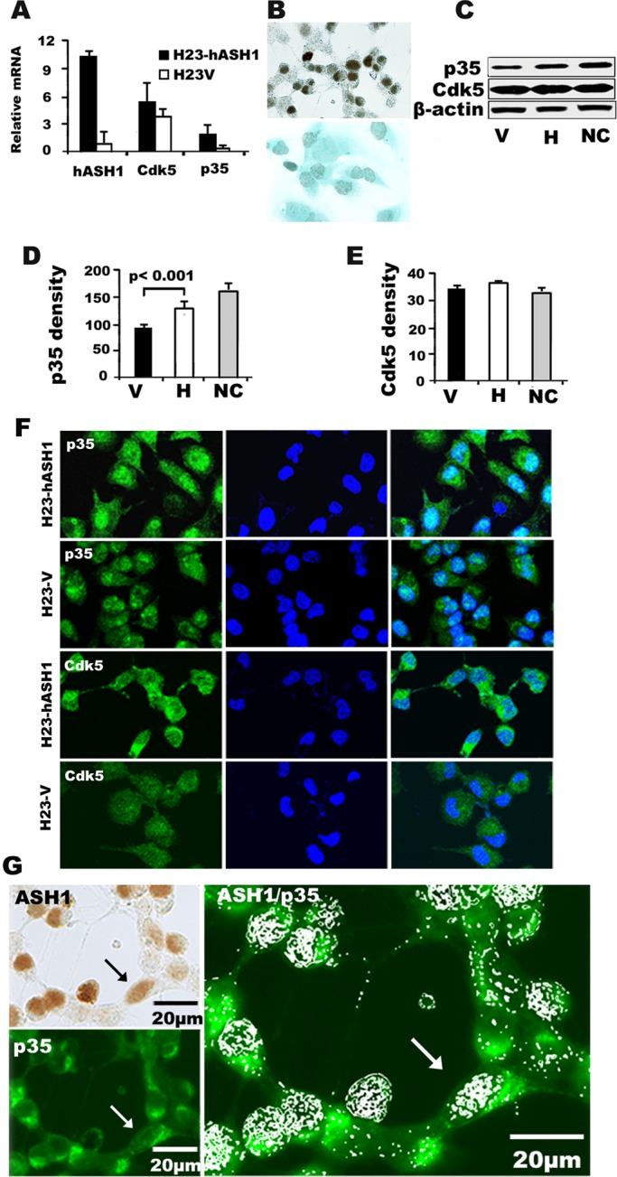
Overexpression of hASH1 in human lung adenocarcinoma cells leads to an increase in p35 expression in vitro. (A) By qRT-PCR, expression of hASH1 and p35 mRNAs was seen to increase in H23 cells transfected with hASH1 (H23-hASH1). Total RNA was extracted from stably expressing H23-hASH1 or control H23 cells (H23-V) that were transfected by vector only. hASH1 expression was increased 10-fold in transfected cells compared with empty vector. hASH1 overexpression significantly increased p35 (p < 0.01) mRNA expression. (B) Intense nuclear hASH1 immunoreactivity in H23-hASH1 cells (top panel). H23-V cells were negative (bottom panel) (immunoperoxidase staining). (C) Total protein lysates from H23- hASH1 (H) or control H23V (V) cells were analyzed by Western blot analysis The levels of p35 and Cdk5 were compared with those seen in NCs from fetal mouse brain, which served as a positive control. (D and E) Quantification of the results in (C) by densitometry showed a significant increase (p < 0.001) in p35 in H compared with V cells. NC, neural cells from embryonic brain. The bars represent means ± SD from three independent experiments. (F) Photomicrographs p35 (green) and Cdk5 (green) expression in H23-hASH1 and H23-V cells by immunofluorescence (left). Nuclei (blue) were counterstained with DAPI (middle). Light blue nuclear staining indicates coexpression (overlay in right). The overall intensity of immunofluorescence was higher in H23-hASH1 cells. (G) Photomicrographs of nuclear expression (brown) of hASH1 in H23-hASH1 cells (top, left: immunoperoxidase staining) and the same view showing p35 (green) expression (bottom, left: immunofluorescence). Colocalization of nuclear hASH1 (pseudowhite grains) and p35 (green) by immunoperoxidase and immunofluorescence, respectively, in H23-hASH1 cells. Black and white arrows point to a typical polarized double-positive cell with cytoplasmic extensions.
