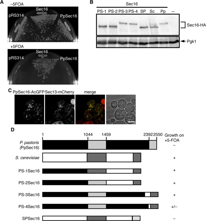
FIGURE 5:
The CTCD is critical for Sec16 function. (A) The ability of PpSec16 to support growth of sec16∆ cells was tested on 5-FOA plates as described in the legend of Figure 1A. (B) Total cell extracts of S. cerevisiae cells (SEY6210) expressing HA-tagged S. cerevisiae (Sc), P. pastoris (Pp), or chimeric Sec16 indicated were separated by SDS–PAGE followed by immunoblotting with anti-HA or anti-Pgk1 antiserum. Chimeras examined are depicted in D. (C) PpSec16 localizes at the ERES in S. cerevisiae cells. S. cerevisiae cells (SEY6210) expressing PpSec16-AcGFP with Sec13-mCherry were grown to mid-log phase and examined by fluorescence microscopy. Scale bar, 4 μm. (D) Chimeras between S. cerevisiae (ScSec16) and P. pastoris (PpSec16) Sec16. A schematic diagram shows chimeric forms of Sec16. The sequence derived from PpSec16 is depicted as black and light gray bars, and the sequence from ScSec16 is shown as white and dark gray bars. Gray bars display the CDC and CTCD. PS-1Sec16; PpSec161-1010 and ScSec16960-2195, PS-2Sec16; PpSec161-1461 and ScSec161423-2195, PS-3Sec16; PpSec161-2266 and ScSec161895-2195, PS-4Sec16; PpSec161-2361 and ScSec161991-2195, SPSec16; and ScSec161-1996 and PpSec162369-2550. The ability of each Sec16 chimera to support the growth of sec16∆ cells was tested on 5-FOA plates as described in the legend of Figure 1A.

