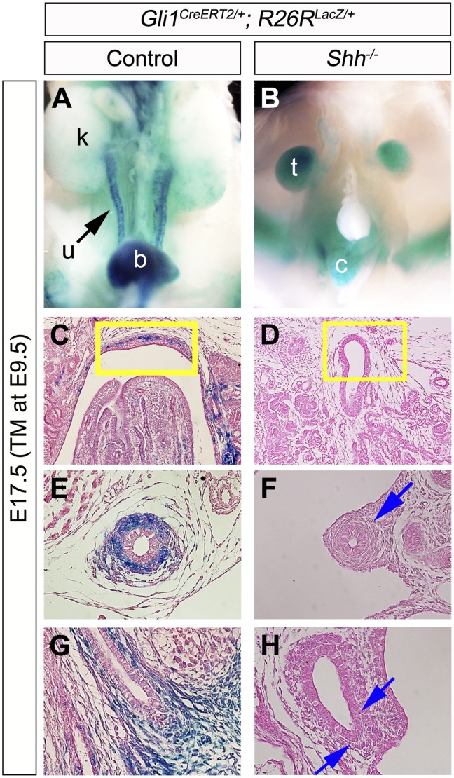
Figure 3. The contribution of Hh-responsive cells in Shh mutant urinary tracts.
Hh-responsive cell contribution assay was performed by utilizing the Gli1CreERT2; R26RLacZ/+ system. Urinary organs of embryos were analyzed at E17.5 following TM treatment at E9.5 (A–H). Gross morphology of X-gal stained embryos (A, B). A weak LacZ signal was detected in Shh−/−; Gli1CreERT2/+; R26RLacZ/+embryos in comparison to control embryos in the renal pelvis (yellow boxes in C, D), the ureter (E, F) and the bladder trigone region (G, H). Blue arrows indicate reduced LacZ signals in the urinary tracts of Shh−/−; Gli1CreERT2/+; R26RLacZ/+ embryos. b: bladder, c: cloaca, k: kidney, t: testis, u: ureter.

