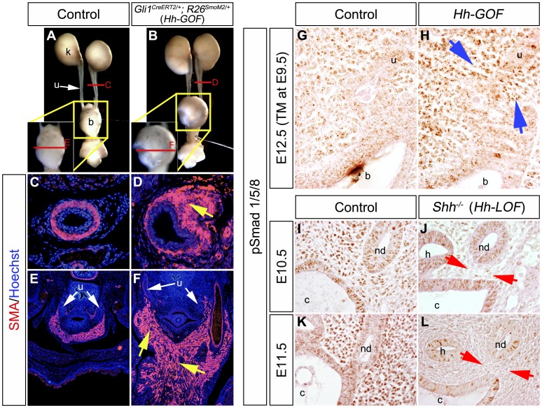
Figure 4. Constitutive activation of Hh signaling led to hyperplasia of urinary smooth muscles and augmented expression of phosphorylated-Smad1/5/8 in the urogenital mesenchyme.
The gain-of-function experiments in Hh-responsive cells were performed by utilizing Gli1CreERT2; R26SmoM2 (Hh-GOF) mice. Gross morphology of urinary tracts in control and mutant embryos at E17.5 after E9.5-TM treatment (A, B). Locations of the magnified bladder regions were indicated by yellow boxes in A and B. Levels of transverse sections in C–F are indicated by red lines in A and B. Immunohistochemistry for the expression of SMA (red) in transverse sections of the ureter (C, D) and the bladder trigone (E, F) regions. Yellow arrows indicate the abnormally expanded urinary smooth muscle layers. Immunohistochemistry for phosphorylated-Smad1/5/8 (pSmad) expression was also performed in Hh-GOF mutants with TM treatment at E9.5 (G, H). The Hh-GOF mutants displayed augmented pSmad expression in peri-cloacal mesenchyme at E12.5 (blue arrows). The Shh−/− (Hh signal loss-of-function mutants: Hh-LOF mutants) displayed a reduction of the pSmad expression at E10.5 and E11.5 (I–L: red arrows). b: bladder, c: cloaca, h: hindgut, nd: nephric duct, k: kidney, u: ureter.

