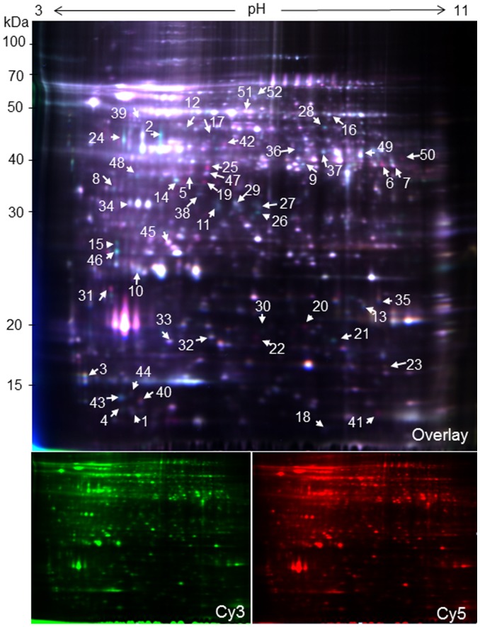
Figure 2. 2D-DIGE images of anther proteins from the WT and male sterile mutant YX-1 in wolfberry.
Extracts from the WT and YX-1 anther of three independent biological repeat experiments were differentially labeled with the spectrally resolvable CyDye fluors Cy3 and Cy5 and separated by two-dimensional electrophoresis (2-DE) on 13-cm (pH 3–11) IPG strips and 12.5% polyacrylamide gels. A merged image of Cy5-labeled YX-1 (red) and Cy3-labeled WT (green) is shown. Arrowed and numbered spots in the image are differentially expressed protein spots. Molecular markers (in kDa) are shown on the left.

