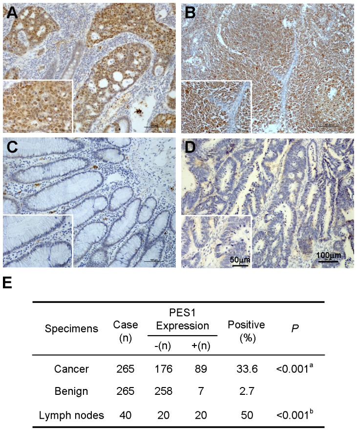
Figure 1. PES1 is overexpressed in colon cancer.

Immunohistochemical analysis of PES1 expression in human colon cancer tissues. The figures show the strongly nuclei staining of PES1 in colon cancer tissues (A) and lymph nodes (B). Negative staining of PES1 in non-cancerous tissues adjacent to tumor was shown in (C). Normal mouse IgG was used as a negative control in colon cancer tissues and shown in (D). Representative low (×100) and high (×400) magnification are shown. (E) Summary of PES1 expression in human colon cancer tissues, matched adjacent tissues, and lymph nodes. a indicates significant difference between colon cancer versus matched adjacent tissue. b indicates significant difference between lymph nodes versus adjacent noncancerous tissues.
