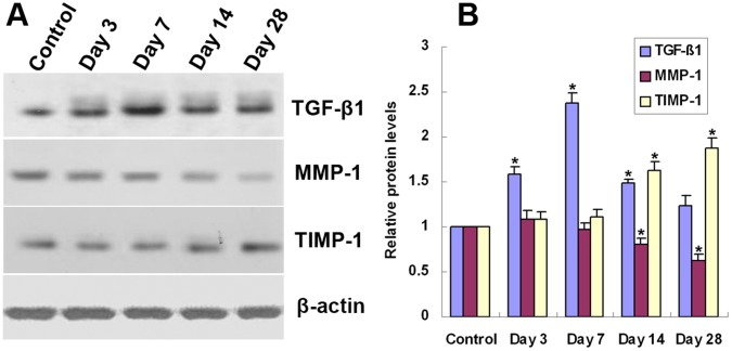
Figure 4. Temporal expression of TGF-β1, MMP-1, and TIMP-1 protein in grafted veins.
Western blot was performed to determine the expression of TGF-β1, MMP-1 and TIMP-1 in grafted veins on days 3, 7, 14, and 28. β-actin was used as a loading control. (A) Representative Western blot image. (B) Quantitative analysis of Western blot images (n = 6). *P<0.05 vs. Control.

