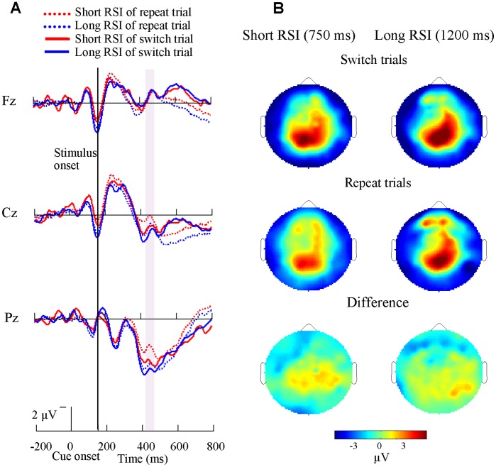
Figure 5. Cue-related ERP waveforms and brain topographies for short CSI conditions.
(A) Grand-average cue-related ERP waveforms at midline sites (frontal, Fz, central, Cz, and parietal, Pz) are shown for the 14 subjects in the short CSI conditions across both repeat and switch trials. A large sharp parietal positivity emerged at about 400 ms after cue onset. Switch trials evoked larger amplitude than that of repeat trials. (B) Brain topographies in the short CSI conditions across repeat (mid) and switch trials (top) and the difference topographies between switch and repeat trials (bottom) are shown. There was more activation for switch trials in parietal cortex.

