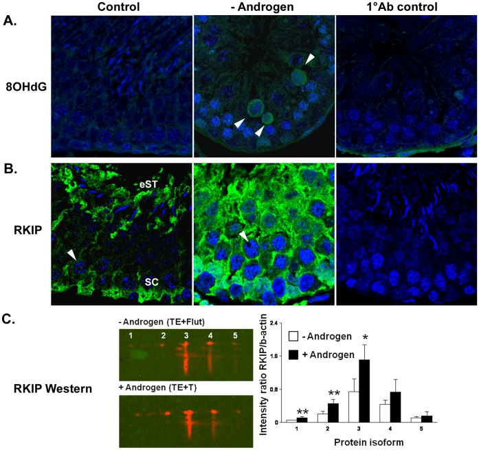
Figure 3. Localisation of oxidised DNA adducts and RKIP during androgen manipulation in vivo.
A. Immunohistochemical localisation of oxidised DNA adducts as detected by 8OHdG (green) labelling in testis from Control and - Androgen (androgen suppressed, TE+Flutamide) rats. A negative control for the primary antibody is also shown (1°Ab Control) in a TE+Flutamide-treated testis. Positively labelled pachytene spermatocytes were only apparent during androgen suppression (arrowheads). B. Immunohistochemical localisation of RKIP (green) in testis from Control and - Androgen (androgen suppressed, TE+Flutamide). A negative control for the primary antibody is also shown (1°Ab Control). In controls, staining was most apparent in Sertoli cells (SC) and elongating spermatid cytoplasm (eST), but was faintly present in pachytene spermatocyte cytoplasm (arrowheads). During androgen suppression (- Androgen) a marked increase in immunostaining for RKIP was noted throughout the epithelium, with cytoplasmic staining more obvious in pachytene spermatocytes (arrowhead). In A and B, nuclei are labelled blue (TOPRO). C. Confirmation of changes in expression of androgen-responsive RKIP isoforms; the left panel shows representative images of the 2D-Western during androgen blockade (-Androgen, TE+Flutamide) compared to androgen replacement (+ Androgen, TE+T24). Blots were performed on pooled samples from the same individual animals used for the 2D-DIGE proteomics. Five distinct isoforms (#1– # 5) were resolved. Results of the densitometric analysis (right panel) from 2D western blots revealed that 3 isoforms showed significant (* p<0.05, ** p<0.01) differences between the –Androgen and + Androgen groups. Data is shown as mean ± SD (n = 4 separate experiments).

