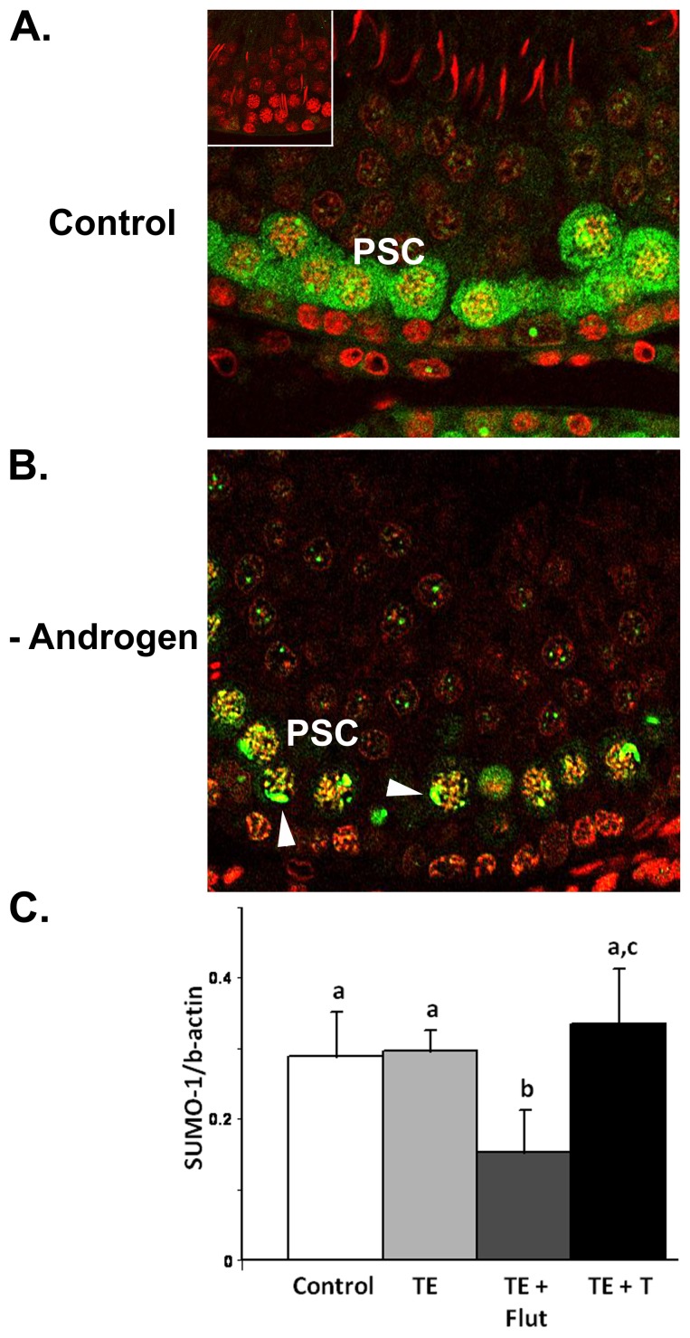
Figure 5. SUMO1 during androgen manipulation.
A. Immunohistochemical localisation of SUMO1 (green) in control testis. Staining is observed in the cytoplasm of pachytene spermatocytes (PSC) in this stage VII tubule, whereas no staining was observed in the primary antibody control (inset). Cell nuclei were visualized with TOPRO (red). B. SUMO1 immunostaining in pachytene spermatocyte (PSC) cytoplasm was reduced during androgen suppression, however staining associated with the nuclei and the XY body (arrowheads) was preserved. C. Densitometric analysis of 15 kDa SUMO1 (i.e. ‘free’ SUMO1) in 1D-western blots with n = 4 separate animals/group from the four different treatments. Different letters denote statistical differences (p<0.01) between groups. During androgen blockade (TE+Flut), there was a significant decrease in free SUMO1 compared to control. Data is shown as mean ± SD.

