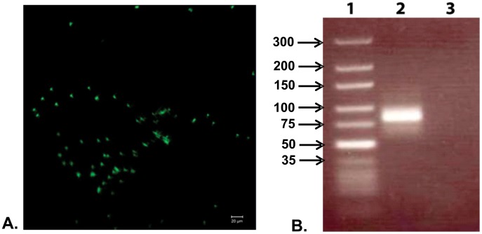
Figure 2. One-step selection and amplification of the aptamer candidates.
A. The observed fluorescence on the toxin immobilized glass coverslip after washing. The glass coverslip was washed with extensive amounts of binding buffer, effectively removing all nonspecific adhesion to the coverslip. The pattern of highly localized fluorescent dots and the absence of background smear provide an indication of successful selection; B. PCR amplification of the eluted bound sequence. Lane 1: Marker DNA, lane 2: Amplified product from the eluted DNA aptamers, lane 3: PCR amplification without using a template DNA (negative control).

