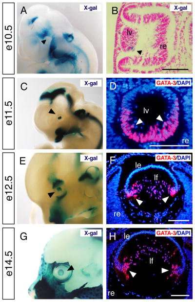
Fig. 1. Optic expression of GATA-3 during embryogenesis.
A–F: Gata3lacZ knock-in heterozygotes at e10.5, e11.5, e12.5 and e14.5 were stained with X-gal and photographed in whole-mounts (A, C, E and G) and section (B). Black arrowheads in each panel indicate lacZ-positive lens. GATA-3 immunoreactivity was specifically observed in the posterior part of the lens vesicle in e11.5 embryos and in lens fiber cells from e12.5 onward (white arrowheads in D, F and H). le, lens epithelium; lf, lens fiber; lv, lens vesicle; re, retina. Scale bars = 100 μm.

