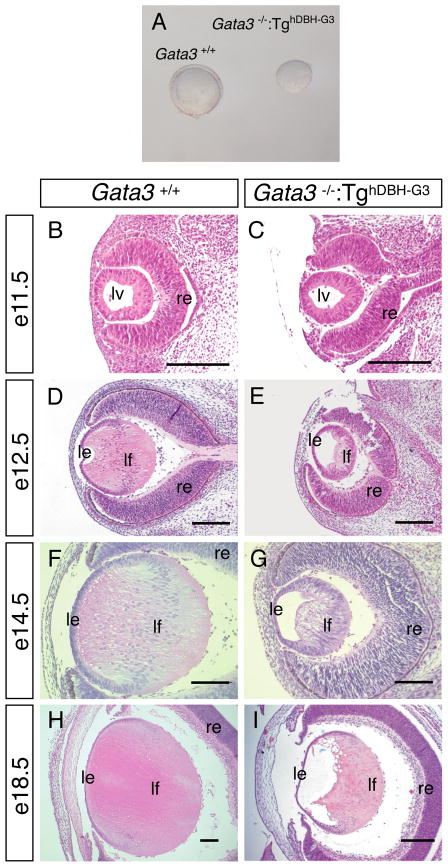
Figure 2. Defective lens development in rescued Gata3−/−:TghDBH-G3 embryos.
A: Lenses dissected from e16.5 Gata3−/−:TghDBH-G3 embryos are smaller than wild type. B–I: Sagittal sections of e11.5 (B, C), e12.5 (D, E), e14.5 (F, G) and e18.5 (H, I) Gata3+/+ and Gata3−/−:TghDBH-G3 embryos were stained with hematoxylin and eosin. In e11.5 Gata3 mutant embryos, the lens vesicle appeared to be normal when compared to wild type (B and C). From e12.5 onward, GATA-3-deficient lenses were smaller and contained a visible lumen as the lens fiber cells failed to elongate to fill the cavity during late embryogenesis. Note that the lens fiber cells of e18.5 Gata3−/−:TghDBH-G3 embryos remained highly nucleated (I) in contrast to the wild type lens fiber cells (H). lv, lens vesicle, le, lens epithelium; lf, lens fiber; re, retina. Scale bars = 100 μm.

