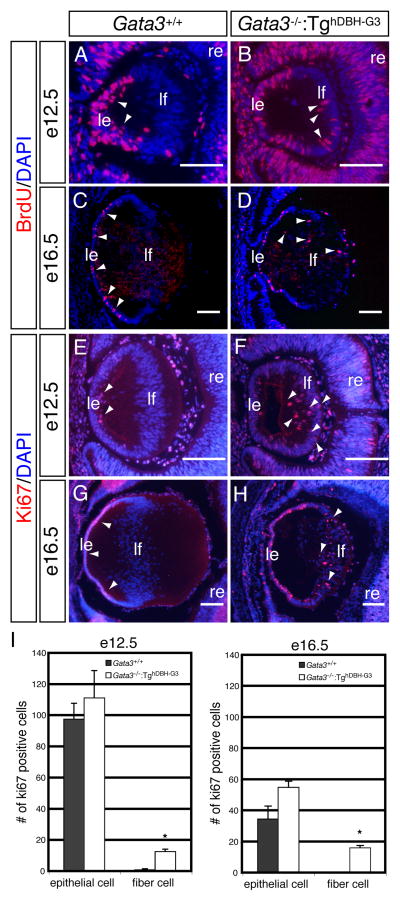
Figure 5. Altered cellular proliferation in the lenses of Gata3−/−:TghDBH-G3 embryos.
A–H: In wild type embryos, Ki67- or BrdU-immunoreactive cells were observed exclusively in the anterior lens epithelium (arrowheads in A, C, E and G), whereas an increased number of mitotic cells were present in the lens fiber cells located in the lens posterior (arrowheads in B, D, F and H) in e12.5 and e16.5 Gata3−/−:TghDBH-G3 embryos. le, lens epithelium; lf, lens fiber; re, retina. Scale bars: 100 μm. I: Quantification of ki-67-positive cells in e12.5 and e16.5 embryonic lenses epithelial and fiber cells from wild type (n=6) and Gata3−/−:TghDBH-G3 (n=6) embryos (*P<0.05; Student’s t-test). Six lenses from six different embryos of each genotype were analyzed. Data are presented as means±s.e.m. The statistical significance of the differences between Gata3−/−:TghDBH-G3 and Gata3+/+ are indicated by (*P<0.05; Student’s t-test).

