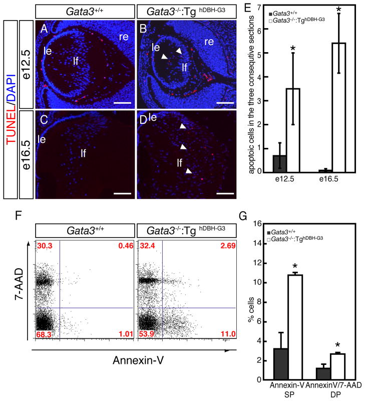
Figure 6. Increased cell death in lens fiber cells of Gata3−/−:TghDBH-G3 mutant embryos.
A–D: TUNEL assays detected an increase in the number of apoptotic cells in the posterior chamber of e12.5 and e16.5 Gata3−/−:TghDBH-G3 lenses (arrowheads). le, lens epithelium; lf, lens fiber; re, retina. Scale bars: 100 μm. E: Quantification of TUNEL-positive cells in e12.5 and e16.5 embryonic lenses of wild type (n=6) and Gata3−/−:TghDBH-G3 (n=6) embryos. Six lenses from six different embryos of each genotype were analyzed. Data are presented as means±s.e.m. The statistical significance of the differences between Gata3−/−:TghDBH-G3 and Gata3+/+ are indicated by (*P<0.05; Student’s t-test). F: Representative flow cytometric profiles of single-cell suspensions that were dissociated from lenses of e18.5 wild type or Gata3−/−:TghDBH-G3 embryos, stained with PE-conjugated Annexin-V antibody (horizontal axis) and 7-AAD (vertical axis). The percentage of cells in each quadrant is indicated. G: In the lens fiber cells from e18.5 Gata3−/−:TghDBH-G3 embryos, the Annexin-V-single positive (SP) population, representing early apoptotic cells, increased by more than three-fold (10.85±0.23% in Gata3 mutant [n=5], 3.12±1.23% in wild type control [n=5]). The 7-AAD- and Annexin-V-double positive (DP) cell population (representing late apoptotic and necrotic cells) also increased by more than 2-fold (1.06±0.23% in Gata3 mutant [n=5], 3.09±0.06% in wild type control [n=5]).

